


Bax Recombinant Rabbit Monoclonal Antibody [SZ3-07]
Recombinant Rabbit monoclonal Antibody
Synthetic peptide within Human Bax aa 1-50 / 192.
Human, Mouse, Rat (Predicted: Goat)
WB, IF-Cell, FC
Predicted band size: 21 kDa
HeLa cell lysate, MCF7 cell lysate, HEK-293 cell lysate, bEnd.3 cell lysate, C2C12 cell lysate, PC-12 cell lysate, C6 cell lysate, C2C12, HeLa.
unconjugated
SZ3-07
Liquid
1ug/ul
Store at +4℃ after thawing. Aliquot store at -20℃ or -80℃. Avoid repeated freeze / thaw cycles.
1*TBS (pH7.4), 0.05% BSA, 40% Glycerol. Preservative: 0.05% Sodium Azide.
IgG
Protein A affinity purified.
WB
1:20,000-1:50,000
IF-Cell
1:100
FC
1:1,000
| Human | 查看 50 篇文献如下 |
| Mouse | 查看 39 篇文献如下 |
| Rat | 查看 15 篇文献如下 |
| Pig | 查看 5 篇文献如下 |
| Chicken | 查看 2 篇文献如下 |
| rat | 查看 2 篇文献如下 |
| Silkworm | 查看 1 篇文献如下 |
| Channel Catfish | 查看 1 篇文献如下 |
| Ictalurus punctatus | 查看 1 篇文献如下 |
| mice | 查看 1 篇文献如下 |
| Rats | 查看 1 篇文献如下 |
In healthy mammalian cells, the majority of BAX is found in the cytosol, but upon initiation of apoptotic signaling, Bax undergoes a conformational shift. Upon induction of apoptosis, BAX becomes organelle membrane-associated, and in particular, mitochondrial membrane associated. BAX is believed to interact with, and induce the opening of the mitochondrial voltage-dependent anion channel, VDAC. Alternatively, growing evidence also suggests that activated BAX and/or Bak form an oligomeric pore, MAC in the MOM (mitochondrial outer membrane). This results in the release of cytochrome c and other pro-apoptotic factors from the mitochondria, often referred to as mitochondrial outer membrane permeabilization, leading to activation of caspases. This defines a direct role for BAX in mitochondrial outer membrane permeabilization. BAX activation is stimulated by various abiotic factors, including heat, hydrogen peroxide, low or high pH, and mitochondrial membrane remodeling. In addition, it can become activated by binding BCL-2, as well as non-BCL-2 proteins such as p53 and Bif-1. Conversely, BAX can become inactivated by interacting with VDAC2, Pin1, and IBRDC2.
1. He G et al. Gadd45b prevents autophagy and apoptosis against rat cerebral neuron oxygen-glucose deprivation/reperfusion injury. Apoptosis 21:390-403 (2016).
2. Chen B et al. Inhibition of miR-29c promotes proliferation, and inhibits apoptosis and differentiation in P19 embryonic carcinoma cells. Mol Med Rep 13:2527-35 (2016).
Belongs to the Bcl-2 family.
Expressed in a wide variety of tissues. Isoform Psi is found in glial tumors. Isoform Alpha is expressed in spleen, breast, ovary, testis, colon and brain, and at low levels in skin and lung. Isoform Sigma is expressed in spleen, breast, ovary, testis, lung, colon, brain and at low levels in skin. Isoform Alpha and isoform Sigma are expressed in pro-myelocytic leukemia, histiocytic lymphoma, Burkitt's lymphoma, T-cell lymphoma, lymphoblastic leukemia, breast adenocarcinoma, ovary adenocarcinoma, prostate carcinoma, prostate adenocarcinoma, lung carcinoma, epidermoid carcinoma, small cell lung carcinoma and colon adenocarcinoma cell lines.
Mitochondrion membrane, Cytoplasm.
Apoptosis regulator BAX antibody
BAX antibody
Bax-protein antibody
BAX_HUMAN antibody
BAXA antibody
Baxdelta2G9 antibody
Baxdelta2G9omega antibody
Baxdelta2omega antibody
Bcl-2-like protein 4 antibody
BCL2 associated X protein antibody
展开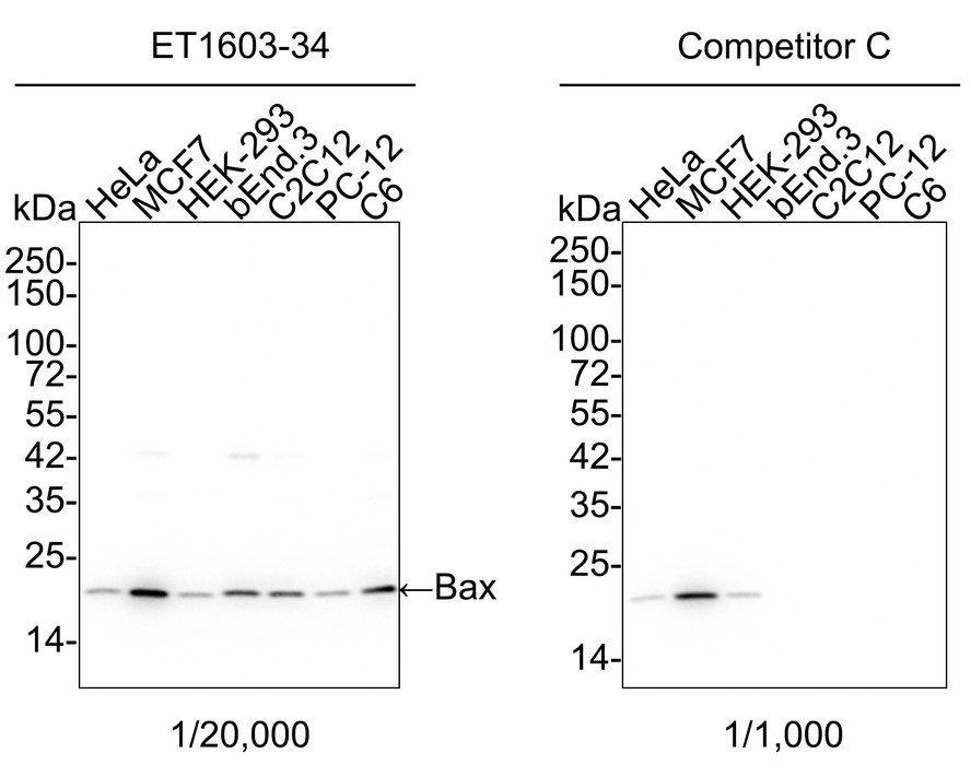
Western blot analysis of Bax on different lysates with Rabbit anti-Bax antibody (ET1603-34) at 1/20,000 dilution and competitor's antibody at 1/1,000 dilution.
Lane 1: HeLa cell lysate
Lane 2: MCF7 cell lysate
Lane 3: HEK-293 cell lysate
Lane 4: bEnd.3 cell lysate
Lane 5: C2C12 cell lysate
Lane 6: PC-12 cell lysate
Lane 7: C6 cell lysate
Lysates/proteins at 15 µg/Lane.
Predicted band size: 21 kDa
Observed band size: 21 kDa
Exposure time: 30 seconds; ECL: K1801;
4-20% SDS-PAGE gel.
Proteins were transferred to a PVDF membrane and blocked with 5% NFDM/TBST for 1 hour at room temperature. The primary antibody (ET1603-34) at 1/20,000 dilution and competitor's antibody at 1/1,000 dilution were used in 5% NFDM/TBST at 4℃ overnight. Goat Anti-Rabbit IgG - HRP Secondary Antibody (HA1001) at 1/50,000 dilution was used for 1 hour at room temperature.
![<span style="font-weight: bold;">☑ Knockout (KO)</span><br /><br />All lanes: Western blot analysis of Bax with anti-Bax antibody [SZ3-07] (<a href="/products/ET1603-34" style="font-weight: bold;text-decoration: underline;">ET1603-34</a>) at 1/1,000 dilution.<br />Lane 1/2: Wild-type Hela whole cell lysate (20 µg).<br />Lane 3/4: Bax knockout Hela whole cell lysate (20 µg).<br /><br /><a href="/products/ET1603-34" style="font-weight: bold;text-decoration: underline;">ET1603-34</a> was shown to specifically react with Bax in wild-type Hela cells. No band was observed when Bax knockout samples were tested. Wild-type and Bax knockout samples were subjected to SDS-PAGE. Proteins were transferred to a PVDF membrane and blocked with 5% NFDM in TBST for 1 hour at room temperature. The primary antibody (<a href="/products/ET1603-34" style="font-weight: bold;text-decoration: underline;">ET1603-34</a>, 1/1,000) and Loading control antibody (Rabbit anti-β-actin, <a href="/products/R1207-1" style="font-weight: bold;text-decoration: underline;">R1207-1</a>, 1/1,000)was used in 5% BSA at room temperature for 2 hours. Goat Anti-Rabbit IgG-HRP Secondary Antibody (<a href="/products/HA1001" style="font-weight: bold;text-decoration: underline;">HA1001</a>) at 1:200,000 dilution was used for 1 hour at room temperature.](https://storage.huabio.cn/huabio/productImg/ET1603-34_2.jpg?v=20241015165532)
☑ Knockout (KO)
All lanes: Western blot analysis of Bax with anti-Bax antibody [SZ3-07] (ET1603-34) at 1/1,000 dilution.
Lane 1/2: Wild-type Hela whole cell lysate (20 µg).
Lane 3/4: Bax knockout Hela whole cell lysate (20 µg).
ET1603-34 was shown to specifically react with Bax in wild-type Hela cells. No band was observed when Bax knockout samples were tested. Wild-type and Bax knockout samples were subjected to SDS-PAGE. Proteins were transferred to a PVDF membrane and blocked with 5% NFDM in TBST for 1 hour at room temperature. The primary antibody (ET1603-34, 1/1,000) and Loading control antibody (Rabbit anti-β-actin, R1207-1, 1/1,000)was used in 5% BSA at room temperature for 2 hours. Goat Anti-Rabbit IgG-HRP Secondary Antibody (HA1001) at 1:200,000 dilution was used for 1 hour at room temperature.

Immunocytochemistry analysis of C2C12 cells labeling Bax with Rabbit anti-Bax antibody (ET1603-34) at 1/100 dilution.
Cells were fixed in 4% paraformaldehyde for 20 minutes at room temperature, permeabilized with 0.1% Triton X-100 in PBS for 5 minutes at room temperature, then blocked with 1% BSA in 10% negative goat serum for 1 hour at room temperature. Cells were then incubated with Rabbit anti-Bax antibody (ET1603-34) at 1/100 dilution in 1% BSA in PBST overnight at 4 ℃. Goat Anti-Rabbit IgG H&L (iFluor™ 488, HA1121) was used as the secondary antibody at 1/1,000 dilution. PBS instead of the primary antibody was used as the secondary antibody only control. Nuclear DNA was labelled in blue with DAPI.
Beta tubulin (M1305-2, red) was stained at 1/100 dilution overnight at +4℃. Goat Anti-Mouse IgG H&L (iFluor™ 594, HA1126) was used as the secondary antibody at 1/1,000 dilution.
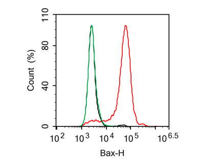
Flow cytometric analysis of HeLa cells labeling Bax.
Cells were fixed and permeabilized. Then stained with the primary antibody (ET1603-34, 1μg/mL) (red) compared with Rabbit IgG Isotype Control (green). After incubation of the primary antibody at +4℃ for an hour, the cells were stained with a iFluor™ 488 conjugate-Goat anti-Rabbit IgG Secondary antibody (HA1121) at 1/1,000 dilution for 30 minutes at +4℃. Unlabelled sample was used as a control (cells without incubation with primary antibody; black).

期刊: Cell Communication And Signaling
应用: WB
反应种属: Mouse
发表时间: 2025 Mar


抗体说明书Product Type中有详细描述抗体种类,“monoclonal”即为单克隆抗体,“polyclonal”为多克隆抗体。Huabio产品详情页说明中有直接写明抗体类型。如果对抗体的特异性要求高,用量较大或需要长期使用一致的抗体,可以选择单克隆抗体。多克隆抗体即使使用相同的抗原制备,不同批次间也会存在差异,因而在特异性、一致性方面有很大的局限。多抗识别多个抗原表位,即使是有少数几个抗原表位被破坏或者抗原构象改变,实验的结果也不会受到影响,因此当实验种属未经过验证时也可以选择多抗。若对抗体的特异性要求不高,需要做沉淀和凝集反应的检测性实验或者只需做ELISA检测,可以选择多克隆抗体。
Huabio抗体的储存条件见产品网页存放说明或说明书中“Storage Instruction”部分。我们的大部分非偶联抗体应储存于-20℃。因为Huabio大多数非偶联抗体是在甘油中的,抗体在此温度下不会冻住,但是也有部分抗体未加甘油,需要储存在4℃。有关特定产品存储建议的信息,请务必参阅产品网页或说明书。
抗体名称括号中的数字和字母为克隆号。克隆号在历史上被用于区分不同的单克隆抗体。如果两个抗体有同样的克隆号,那么它们是来源于同一个单克隆的抗体,但它们可能在种属反应和应用上有差异。


